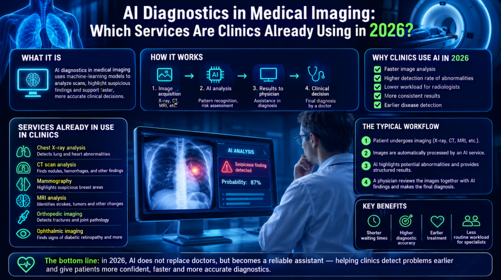
AI-based medical imaging has moved beyond experimental research and is increasingly becoming part of routine clinical workflows. Algorithms can already assist with the analysis of X-rays, CT scans, mammograms, MRI studies, and other medical images, helping specialists identify suspicious findings and prioritize examinations. For patients, however, it is important to understand that AI generally supports clinical decision-making rather than replacing the physician responsible for the final diagnosis.
What Is AI-Based Medical Imaging Diagnostics and How Does It Work?
AI imaging systems use machine-learning models to identify patterns in medical images and highlight findings that may require a specialist’s attention.
Most modern systems are based on machine learning and deep neural networks trained on large collections of medical images. During training, an algorithm learns statistical patterns associated with particular anatomical structures, abnormalities, or diseases. When a new examination is processed, the system compares its visual characteristics with patterns learned from the training data.
Depending on its purpose, an AI system may detect a suspected abnormality, outline a region of interest, measure anatomical structures, classify findings, calculate quantitative parameters, or assign a probability score. Some solutions can also compare current and previous examinations or help prioritize studies that may contain urgent findings.
For example, an algorithm processing a chest CT scan may highlight pulmonary nodules or other suspicious areas for a radiologist to review. A mammography system may mark regions associated with possible breast abnormalities, while an X-ray algorithm may draw attention to findings compatible with fractures or lung disease.
The crucial distinction is that detecting an imaging feature is not necessarily the same as making a clinical diagnosis. A physician can consider symptoms, medical history, laboratory results, previous examinations, medications, and other information that may not be available to the imaging algorithm.
Which AI Services Are Already Used in Russian Clinics?
Russian healthcare organizations use AI solutions in several areas of radiology and medical imaging, although the exact systems available depend on the region, clinic, infrastructure, and regulatory status of individual products.
One of the most visible examples is Moscow’s large-scale experimentation and implementation of computer-vision technologies in radiology. AI services have been integrated into imaging workflows to assist specialists with analysis and prioritization across multiple types of examinations.
The Russian market also includes domestic developers offering software designed for radiology departments, screening programs, and clinical decision support. Rather than treating every AI product as interchangeable, it is more useful to distinguish them by their clinical task:
- Chest X-ray analysis — algorithms can help identify and highlight findings associated with pulmonary abnormalities and other conditions visible on radiographs.
- Chest CT analysis — computer-vision systems can assist with detecting pulmonary nodules, evaluating lung abnormalities, and performing quantitative measurements.
- Mammography — AI can mark suspicious areas and provide an additional layer of analysis during breast cancer screening.
- Neurological imaging — some systems process CT or MRI examinations to identify findings that may require rapid assessment, including abnormalities associated with acute neurological conditions.
- Automated measurements and image triage — AI can calculate parameters, identify potentially urgent examinations, and help radiologists organize their worklists more efficiently.
The presence of AI in a clinic does not automatically mean that an algorithm independently issues a diagnosis. Its role can range from background image processing and measurements to providing a structured second opinion for the radiologist.
Which Types of Medical Images Does AI Analyze Most Often?
AI is particularly well suited to standardized digital imaging modalities in which large numbers of examinations can be processed using repeatable visual criteria.
X-rays are one of the most natural applications. They are widely performed, relatively standardized, and frequently used for screening and initial assessment. Algorithms can analyze chest radiographs, skeletal images, and other studies depending on the intended purpose of a particular system.
Computed tomography is another major area. A single CT examination can contain hundreds or even thousands of images, creating a substantial workload for radiologists. AI can search these datasets for predefined abnormalities, perform measurements, segment organs or lesions, and highlight regions requiring closer inspection.
Mammography is also frequently associated with AI-assisted analysis because screening involves large numbers of relatively standardized examinations. An algorithm can act as an additional reader by marking potentially suspicious regions for assessment by a specialist.
MRI presents additional opportunities but is technically more complex because examination protocols, sequences, anatomical regions, and acquisition parameters can vary considerably. Nevertheless, AI is increasingly used for segmentation, quantitative analysis, image reconstruction, and detection or classification tasks in selected applications.
AI can also work with other forms of digital medical imagery, including ophthalmic images and certain ultrasound applications. Its usefulness depends not simply on the imaging modality but on whether the system has been developed, validated, and authorized for the specific clinical task.
How Is an Image Checked with AI During a Medical Appointment?
In a typical workflow, AI processes the examination alongside the clinic’s existing imaging systems and provides its results to a medical professional for interpretation.
For patients, the process may look almost identical to an examination performed without AI. Much of the automated analysis happens within the clinic’s digital infrastructure.
- The examination is performed. The patient undergoes an X-ray, CT, MRI, mammography, or another imaging procedure according to the prescribed protocol.
- The images enter the clinic’s information system. If an appropriate AI service is integrated into the workflow, a copy or relevant imaging data can be automatically submitted for algorithmic analysis.
- The AI processes the study. Depending on its function, the system may highlight suspicious regions, calculate measurements, classify findings, generate probability estimates, or produce a structured analytical output.
- A physician reviews the examination. The specialist evaluates the original images together with available AI results and relevant clinical information. The physician then prepares the medical report or determines whether further assessment is necessary.
AI therefore usually operates as another source of information within the diagnostic process. A specialist should still evaluate whether the algorithm’s output corresponds to what is actually visible on the images and whether it is clinically meaningful for the particular patient.
Limitations and Responsibility for the Diagnosis
AI can improve the efficiency and consistency of image analysis, but its output remains subject to technical limitations, clinical context, and professional medical interpretation.
No diagnostic algorithm has perfect sensitivity and specificity. False-positive results are possible, meaning the system identifies a suspicious finding that is not clinically significant. False-negative results can also occur when an abnormality is not detected.
Performance may be affected by image quality, unusual anatomy, acquisition parameters, artifacts, previous surgery, implants, rare diseases, or differences between the patient population being examined and the datasets used to develop and validate the model.
Another limitation is context. An imaging algorithm may analyze a CT scan extremely quickly but still have less information than the treating physician. The significance of the same radiological finding can differ depending on a patient’s age, symptoms, previous examinations, laboratory results, medical history, and treatment.
For this reason, patients should not interpret an AI-generated score, marker, or highlighted area as an independent definitive diagnosis. Questions about the meaning of imaging findings and subsequent treatment should be discussed with a qualified healthcare professional.
Responsibility is also an important issue for clinics. Healthcare organizations need to define how AI output is incorporated into their clinical workflow, who reviews it, how disagreements between the algorithm and physician are handled, and how results are documented. Applicable medical-device, healthcare, privacy, and data-processing requirements must also be considered.
How Clinics Can Implement AI Diagnostics and What to Consider
Successful AI implementation requires more than purchasing software: a clinic needs a clearly defined clinical task, validated technology, reliable integration, data protection, and a process for monitoring performance after deployment.
The first step is to identify a specific problem. A radiology department may want to reduce the time spent on routine measurements, prioritize potentially urgent examinations, provide radiologists with an additional detection tool, or increase throughput in a screening program. A measurable objective makes it easier to determine whether an AI system provides practical value.
The clinic should then evaluate the solution’s intended medical purpose and regulatory status. Documentation should clearly describe which imaging modalities, anatomical regions, findings, and patient populations the system is designed to analyze. Evidence supporting performance should also be relevant to the clinic’s actual use case.
Technical integration deserves equal attention. The AI service may need to interact with PACS, RIS, electronic medical records, or other healthcare information systems. Administrators should understand where images are processed, how quickly results are returned, how failures are handled, and whether clinicians can continue working normally when the AI service is unavailable.
Medical data protection is another essential requirement. Clinics should evaluate how patient information is transmitted, stored, accessed, logged, and protected, particularly when processing involves external infrastructure.
Before full-scale deployment, a controlled pilot can help determine how the technology performs in the clinic’s own environment. Useful indicators may include processing time, false-positive and false-negative cases, agreement with specialists, changes in reporting time, and the frequency with which AI findings affect clinical interpretation.
Finally, AI performance should be monitored after implementation. Imaging equipment, clinical protocols, patient populations, software versions, and workflows can change over time. Regular quality assessment helps ensure that an algorithm remains a useful clinical instrument rather than becoming an automated step that staff follow without sufficient scrutiny.
The most effective model is therefore not “AI instead of a doctor,” but AI integrated into a well-designed medical workflow. When the responsibilities of the algorithm and the healthcare professional are clearly separated, automated image analysis can help specialists manage large volumes of information while preserving human oversight of decisions that affect patient care.